
Are you scheduled for a renal ultrasound and wondering what it is all about? Or perhaps your doctor has recommended one, but you’re not quite sure what to expect. A renal ultrasound is a non-invasive imaging test that uses high-frequency sound waves to create detailed images of the kidneys and surrounding areas. In this blog post, we’ll break down everything you need to know about renal ultrasounds – from their benefits and types, to how to prepare for one and what to expect during the procedure. So grab a cup of coffee (or tea!) and let’s decode the results of your upcoming renal ultrasound!
What is a renal ultrasound?
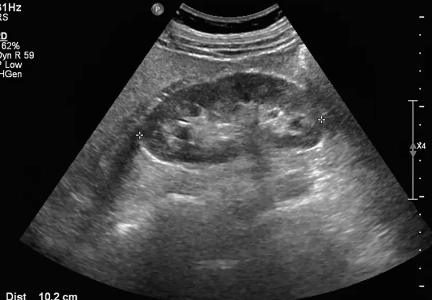
A renal ultrasound is a non-invasive imaging test that uses high-frequency sound waves to produce images of the kidneys and surrounding areas. Unlike other imaging tests, such as CT scans or MRIs, ultrasounds do not use radiation. Instead, they rely on sound waves that are transmitted through the body and bounce back to create an image.
During a renal ultrasound, a technician will apply a gel-like substance to your skin over the area being studied (usually your abdomen). The technician will then use a small device called a transducer to send sound waves into your body and capture the echoes that bounce back. These echoes are then translated by computer software into detailed images of your kidneys.
Renal ultrasounds can help detect abnormalities in the size or shape of the kidneys, identify cysts or tumors within them, evaluate blood flow to the kidneys, and diagnose conditions like kidney stones or infections. They can also be used to monitor certain kidney conditions over time.
Renal ultrasounds are safe and painless procedures that provide valuable information about kidney health without exposing patients to harmful radiation.
What are the benefits of a renal ultrasound?
A renal ultrasound is a non-invasive diagnostic imaging test that uses sound waves to generate images of the kidneys. This type of imaging can help identify any abnormalities or issues within the kidneys, bladder and ureters. There are several benefits associated with undergoing a renal ultrasound.
Firstly, a renal ultrasound is considered safe as it does not involve exposure to ionizing radiation like other diagnostic tests such as CT scans. This makes it an ideal choice for individuals who require frequent monitoring due to chronic kidney conditions.
Secondly, this procedure provides real-time images which means that doctors can see how well your kidneys are functioning and identify any blockages in real time.
Thirdly, early detection of potential problems through routine screening with ultrasounds can lead to earlier treatment interventions which may prevent further complications from developing.
There are numerous benefits associated with getting a renal ultrasound including its safety profile, ability to provide real-time images and early detection of potential problems. It’s important for individuals at risk for kidney disease or those experiencing symptoms related to the urinary tract system to discuss whether this test may be appropriate for them with their healthcare provider.
Types of renal ultrasounds
There are three types of renal ultrasounds: abdominal, pelvic and transrectal.
An abdominal ultrasound is the most common type of renal ultrasound. This procedure involves placing gel on your abdomen and using a probe to create images of your kidneys. It’s non-invasive and typically takes about 30 minutes.
A pelvic ultrasound may be used in addition to an abdominal ultrasound if further details are needed. Pelvic ultrasounds involve placing the probe on or inside the vagina (for women) or rectum (for men). This allows for more detailed images of the kidneys as well as surrounding organs.
Transrectal ultrasounds are typically only used in men when prostate imaging is necessary. They involve inserting a small probe into the rectum to get closer access to both the prostate gland and kidneys.
It’s important to discuss with your doctor which type(s) of renal ultrasound may be necessary for your individual case, as not everyone will need all three types.
How to prepare for a renal ultrasound
Preparing for a renal ultrasound is relatively easy and straightforward. First, it’s essential to let your doctor know if you have any allergies or medical conditions that could affect the test’s accuracy. They may ask you to avoid certain foods and drinks before the procedure.
Most importantly, it would be best if you drank plenty of water before the test as this helps fill your bladder, allowing for better visualization of the kidneys during the exam. You should also wear loose-fitting clothes so that they are easy to remove or adjust during the scan.
It is crucial not to apply any lotion or oil on your skin around your abdomen area as it can interfere with obtaining clear images of your kidney structures. Be sure to follow all instructions given by your healthcare provider closely; this will help ensure optimal results from the test.
In most cases, there are no after-effects following a renal ultrasound examination; however, some people may experience mild discomfort due to having a full bladder during the process. Remember that preparation plays an important role in ensuring accurate results from a renal ultrasound; thus, follow these simple steps accordingly for optimal outcomes!
What to expect during a renal ultrasound
During a renal ultrasound, you will be asked to lie down on an examination table. The technician will apply gel to the area being examined, which is usually your back or abdomen.
The technician will then use a handheld device called a transducer and move it over the area of your kidney(s) being examined. You may feel some pressure or discomfort as they press down with the transducer.
As the technologist takes images, you may hear whooshing sounds from the equipment used during the exam. This is completely normal and nothing to worry about.
It’s important to stay still throughout the procedure so that clear images can be obtained. The entire process typically takes around 30 minutes depending on how many views are necessary for accurate evaluation.
Afterward, you’ll be able to clean off any remaining gel and get dressed before leaving. Your doctor will review the results of your renal ultrasound with you at a later time.
Undergoing a renal ultrasound is usually painless and non-invasive experience that provides valuable information about your kidney health without exposing you to radiation commonly used in other imaging tests like x-rays or CT scans.
Conclusion
A renal ultrasound is a non-invasive and painless imaging test that uses high-frequency sound waves to create images of the kidneys. It is an essential diagnostic tool in identifying kidney problems or detecting abnormalities early on before they progress into more severe conditions.
The benefits of having a renal ultrasound are numerous; it can help your healthcare provider diagnose and manage various illnesses such as kidney stones, cysts, tumors, infections, and congenital anomalies. By knowing what to expect during the procedure and how to prepare beforehand, you can ensure that your experience goes smoothly.
If you have any concerns or questions regarding renal ultrasounds or your health condition as a whole, it’s always best to consult with your doctor for proper guidance. With regular check-ups and timely screenings like renal ultrasounds, we can take control of our health proactively and stay ahead of any potential issues.