
As women, we know that mammograms are essential for our health. However, when it comes to diagnostic mammograms and ultrasounds, there may be some confusion or anxiety around what they entail. These procedures can detect breast cancer at an early stage, which increases the chances of survival significantly. In this blog post, we’ll explain everything you need to know about diagnostic mammograms and ultrasounds – from how they’re performed to what you should expect before and after the procedure. So sit back, relax, and read on to learn more!
What is a diagnostic mammogram?
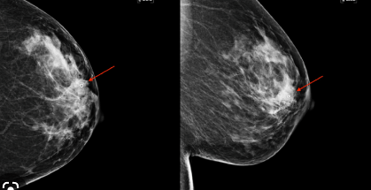
A diagnostic mammogram is a type of X-ray that provides more detailed images of the breast tissue than a screening mammogram. This imaging test is used to evaluate abnormalities found during a screening mammogram or to investigate symptoms such as lumps, pain, nipple discharge, or changes in breast size or shape.
During the procedure, you will be asked to undress from the waist up and wear a hospital gown. The technician will position your breasts on a flat plate and apply another plate from above. The plates will compress your breasts for several seconds while an X-ray machine takes images.
The compression may cause discomfort or slight pain, but it only lasts for a few seconds. Multiple images are taken at different angles to provide complete coverage of each breast.
Afterward, you’ll wait while the technician reviews the quality of the images and decides whether additional pictures are necessary. If they find something suspicious in any area of your breast tissue, they may recommend further testing such as an ultrasound or MRI.
Getting a diagnostic mammogram can help detect potential health concerns early on so that medical professionals can take action quickly and effectively if needed.
What is an ultrasound?
An ultrasound is a diagnostic test that uses high-frequency sound waves to create images of the inside of your body. These images can help doctors diagnose conditions and evaluate injuries without invasive procedures.
During an ultrasound, a technician applies gel to your skin and then moves a small device called a transducer over the area being examined. The transducer emits sound waves that bounce off tissues in your body, creating echoes that are converted into visual images on a computer screen.
Ultrasounds are commonly used during pregnancy to monitor fetal development, but they can also be used to examine organs such as the liver, kidneys or heart. They may also be used for breast imaging when combined with mammography.
One advantage of ultrasounds is that they do not involve radiation exposure like X-rays or CT scans. However, because they rely on sound waves passing through tissue, they may not provide detailed information about structures deep within the body. Your doctor will determine whether an ultrasound is appropriate for your specific condition.
How are they performed?
A diagnostic mammogram involves using low-dose x-rays to obtain images of the breast tissue. The procedure is usually performed in a specialized clinic or hospital by a radiologic technologist.
During the test, you will be asked to undress from your waist up and put on a gown. You will stand in front of an x-ray machine designed specifically for mammography. Your breast will be positioned on a small platform and compressed with another plate.
The compression helps spread out the breast tissue, which makes it easier to capture clear images. You may feel some discomfort during this process, but it should only last for a few seconds.
The technologist will take two views of each breast: one from top to bottom and another from side to side. They may also take additional pictures if they need more information.
An ultrasound uses high-frequency sound waves to produce images of the inside of your body. During an ultrasound exam, you will lie down on an examination table while a technician applies gel over your breasts.
A transducer (a device that emits sound waves) is then moved over your skin capturing images of healthy and abnormal tissues within the breast area. Ultrasound exams are painless and non-invasive procedures that can give doctors valuable insight into any lumps or areas of concern found during other screening tests like mammograms.
Both diagnostic mammograms and ultrasounds are essential tools in detecting potential problems with breast tissue – allowing medical professionals to provide quick results leading towards better treatment options for patients when necessary.
What are the benefits?
One of the biggest benefits of a diagnostic mammogram and ultrasound is early detection. Breast cancer is one of the most common forms of cancer in women, and early detection can make a significant difference in treatment options and outcome. A mammogram can detect small lumps or abnormalities that may be too small to feel during an exam.
Another benefit is peace of mind. For many women, getting regular mammograms can provide reassurance that everything is okay with their breast health. In cases where something abnormal does show up on the test, it allows for prompt action to address any issues.
Because ultrasounds use sound waves instead of radiation, they are considered safe for pregnant women who need further evaluation after an abnormal mammogram result. Ultrasound technology also allows doctors to differentiate between cysts (fluid-filled sacs) and solid masses.
By receiving regular screenings, healthcare providers have a baseline image to compare future images against which increases accuracy over time in detecting potential changes in breast tissue. The benefits outweigh any discomfort or inconvenience associated with these procedures.
Are there any risks?
While diagnostic mammograms and ultrasounds are generally safe procedures, there are some risks associated with them. One of the main concerns is radiation exposure from mammograms, which uses a small amount of ionizing radiation to produce images of the breast tissue.
However, the amount of radiation used in a typical mammogram is very low and unlikely to cause harm. Additionally, modern digital mammography machines use even less radiation than traditional film-based machines.
Ultrasound imaging does not use any ionizing radiation and is considered safe for most people. However, if you have certain medical conditions or are pregnant, your doctor may advise against having an ultrasound.
Another potential risk is false positives or false negatives on the results of these tests. A false positive means that abnormalities were detected but turned out to be benign (non-cancerous), while a false negative means that cancer was missed during the screening process.
It’s important to follow up with additional testing if your doctor suspects something abnormal during either procedure. Always consult with your healthcare provider about any concerns you may have before undergoing any medical test or procedure.
How to prepare for the procedure
Preparing for a diagnostic mammogram and ultrasound is crucial to ensure that the procedure goes smoothly. Firstly, it’s important to inform your doctor if you have any breast symptoms or concerns before scheduling the appointment.
Before the exam, avoid using deodorant, perfume or powders on your chest area as they can interfere with the imaging results. It’s also recommended to wear comfortable clothing that allows easy access to your breasts.
If you’ve had previous mammograms or ultrasounds at a different facility, request copies of those images and bring them along with you to your appointment. This will allow the radiologist to compare them with current images and get a better understanding of any changes in your breast tissue over time.
It’s essential not to schedule this exam during menstruation because hormonal changes can cause breast tenderness and swelling which may affect the accuracy of the test results.
Make sure you communicate openly with your healthcare provider about any concerns or questions you may have regarding preparation for this procedure. They are there to help support and guide you through every step of this process!
What to expect after the procedure
After the procedure of a diagnostic mammogram and ultrasound, patients may experience some mild discomfort or bruising in the area that was examined. This is completely normal and should subside within a few days.
It’s important to avoid strenuous activities or heavy lifting for at least 24 hours after the procedure to allow your body time to recover. Patients are also advised to avoid using any deodorants, lotions, or powders on their breasts before their next appointment.
Results from the exam will typically be available within a week, depending on where it was performed. Your doctor will contact you with the results and discuss any further steps that may need to be taken based on what was found during the examination.
In some cases, additional imaging studies such as an MRI may be recommended if there are any areas of concern detected during your mammogram or ultrasound. However, most women have normal results and can continue with routine breast cancer screening as recommended by their healthcare provider.
It’s important not to worry too much about what comes next after your diagnostic mammogram and ultrasound – simply follow your doctor’s advice and take good care of yourself while you wait for test results!
Conclusion
A diagnostic mammogram and ultrasound are essential tools for detecting breast cancer in its early stages. These procedures are safe, relatively painless, and can provide valuable information about your breast health. By taking the time to prepare properly for your appointment and understanding what to expect during the procedure, you can make sure that you get accurate results that can help guide your treatment plan if necessary.
If you have any concerns about scheduling a diagnostic mammogram or ultrasound, talk to your healthcare provider. They will be able to answer any questions you may have and give you more information about what these tests involve. Remember that early detection is key when it comes to fighting breast cancer, so don’t hesitate to schedule an appointment if recommended by your doctor. Your health is worth it!