
Congratulations, mama-to-be! You have come a long way in your pregnancy journey and are now approaching the exciting milestone of 30 weeks. As you anticipate meeting your little one, it’s only natural to wonder about their development and well-being. This is where ultrasound scans come into play, allowing you to peek inside your womb and see how your baby is growing. In this blog post, we’ll dive deeper into the world of 30 weeks pregnant ultrasounds – what they are, what to expect from them, and how they can benefit both you and your baby. So sit back, relax (as much as possible with that big belly), and let’s explore this fascinating topic together!
What is an ultrasound?
An ultrasound is a medical diagnostic tool that uses high-frequency sound waves to create images of the inside of your body. During pregnancy, ultrasounds are commonly used to check on the development and growth of your baby.
The procedure involves applying gel to your belly and using a wand-like device called a transducer to emit sound waves into your uterus. These sound waves bounce off various structures in your body and return as echoes, which are then translated into visual images on a screen.
Ultrasounds can provide valuable information about the size, position, and health of your baby, as well as detect any potential complications or abnormalities. They can also help determine important factors such as due dates and fetal weight.
While many women may have heard about ultrasounds before becoming pregnant, it’s understandable if you’re feeling nervous or unsure about what exactly happens during the procedure. However, rest assured that ultrasounds are safe for both you and your baby when performed by trained professionals according to recommended guidelines.
How is an ultrasound performed?
Ultrasound is a non-invasive medical test that uses high-frequency sound waves to produce images of the inside of the body. During pregnancy, ultrasounds are used to monitor fetal growth and development.
To perform an ultrasound during pregnancy, you will lie on your back with your abdomen exposed. A special gel will be applied to your belly, which helps conduct the sound waves. The technician will then move a small handheld device called a transducer over your belly, which emits sound waves that bounce off the fetus and return as echoes.
These echoes are picked up by the transducer and sent to a computer, which processes them into images that can be seen on a screen. Depending on how far along in your pregnancy you are, different types of ultrasounds may be performed.
The most common type of ultrasound during pregnancy is called a standard or 2D ultrasound. This produces two-dimensional black-and-white images of the fetus and surrounding structures.
In some cases, doctors may also use 3D or 4D ultrasounds, which provide more detailed images in three dimensions. These can give parents an even clearer view of their baby’s facial features and movements.
Having an ultrasound during pregnancy is safe and painless for both mother and baby. It provides valuable information about fetal development and helps ensure everything is progressing smoothly towards delivery day.
What can you expect to see on an ultrasound during pregnancy?
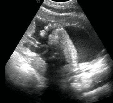
During a 30 weeks pregnant ultrasound, you can expect to see an increasingly developed and more detailed image of your baby. The ultrasound will show the size and position of your baby, as well as their organs, bones, and movements.
The sonographer will measure your baby’s head circumference, abdominal circumference, and femur length. They’ll also check the amount of amniotic fluid surrounding your baby. This information helps determine if your baby is growing normally.
You may be able to see details such as the shape of their nose or ears, fingers and toes or even catch them taking a nap or sucking their thumb! It’s important to note that not all ultrasounds are created equal – some machines may provide clearer images than others.
Another thing that you might notice during this stage is that your little one has become pretty cramped in there! As they grow bigger it becomes harder for them to move around freely which means fewer acrobatics on screen!
A 30 weeks pregnant ultrasound provides valuable information about how both mother and child are progressing through pregnancy.
How can ultrasounds help in the development of the baby?
Ultrasounds play a critical role in monitoring the growth and development of the baby throughout pregnancy. One of the primary benefits of ultrasounds is that they can help detect any potential problems or abnormalities early on, allowing doctors to take necessary actions to ensure a healthy delivery.
During an ultrasound, images are created using high-frequency sound waves that bounce off the baby’s body and are then captured by a device called a transducer. These images provide valuable information about the baby’s size, position, and organ development.
Ultrasounds can also be used to determine important factors such as fetal age, due dates, and gender (if desired). This information helps doctors create personalized care plans for pregnant women based on their individual needs.
In addition to aiding in diagnosis and treatment planning, ultrasounds can also provide parents with an exciting first glimpse at their growing little one. Seeing your baby moving around inside you can be both exhilarating and reassuring during what can sometimes feel like a long journey towards delivery day.
Ultrasounds serve as an essential tool for ensuring the health and well-being of expectant mothers and their babies. By providing detailed insight into fetal development from conception through birth, these tests enable medical professionals to make informed decisions regarding prenatal care while helping parents stay informed every step of the way.
What are some of the risks associated with having an ultrasound during pregnancy?
Ultrasounds are generally considered safe and are routinely used during pregnancy to monitor the growth and development of the baby. However, there are some potential risks associated with having an ultrasound.
One risk is that ultrasounds use high-frequency sound waves which can heat up body tissues. While this is typically not a problem, there is a small chance that it could cause harm to the developing fetus. Additionally, repeated exposure to ultrasounds may increase the risk of low birth weight or preterm labor.
Another concern is that ultrasounds may be misinterpreted or lead to false positives, causing undue stress for expectant parents. In rare cases, an ultrasound may detect a potential issue that leads to unnecessary medical interventions or even termination of a healthy pregnancy.
It’s important to note that these risks are generally very small and most women will have multiple ultrasounds throughout their pregnancy without any complications. If you’re concerned about the safety of ultrasound exams, talk with your healthcare provider about your options and any specific concerns you might have.
Are there any alternatives to having an ultrasound during pregnancy?
While ultrasounds are considered safe during pregnancy, some women may prefer to avoid them or limit their exposure to medical procedures. Fortunately, there are alternative methods for monitoring the health and development of your baby.
One option is fetal doppler monitoring. This involves using a handheld device that emits high-frequency sound waves to detect your baby’s heartbeat. It can be done in the comfort of your own home and does not require any special training.
Another alternative is amniocentesis, which involves taking a small sample of amniotic fluid from around the baby and analyzing it for genetic abnormalities. While this procedure carries more risks than ultrasound, it can provide valuable information about the health of your baby.
Some women choose to rely on regular check-ups with their healthcare provider rather than having additional tests or screenings done. Your doctor or midwife can monitor your pregnancy through physical exams and by listening to your concerns about any symptoms you’re experiencing.
Ultimately, the decision whether or not to have an ultrasound during pregnancy should be based on what feels right for you and your growing family. Be sure to discuss all available options with your healthcare provider before making any decisions.
Conclusion
The 30 weeks pregnant ultrasound is an exciting milestone in a woman’s pregnancy journey. It allows parents to see their baby and monitor its growth and development. With advancements in technology, ultrasounds have become safer and more accurate than ever before.
However, it is important to note that while ultrasounds are generally safe, there are some risks associated with them. Therefore, it is essential to consult with your healthcare provider regarding the frequency of ultrasounds during your pregnancy.
Ultimately, the most critical benefit of having an ultrasound during pregnancy is ensuring the health and well-being of both mother and child. By taking advantage of this amazing technology, you can enjoy a closer look at your growing baby and prepare yourself for the next chapter in your life as a parent.