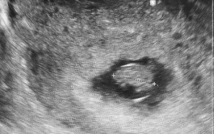
7 Weeks Pregnant Ultrasound: A Guide for Expecting MothersIntroduction

Congratulations! You’re now 7 weeks pregnant and your little bundle of joy is growing rapidly. As an expecting mother, you’re probably feeling a mix of excitement and nerves as you navigate this new chapter in your life. One important milestone during this stage is the 7-week ultrasound, which can give you a sneak peek at your baby’s development. In this guide, we’ll walk you through everything you need to know about the 7-week ultrasound so that you feel confident and prepared for what lies ahead. So sit back, relax and let’s dive into the wonderful world of pregnancy!
What is an Ultrasound?
An ultrasound is a noninvasive medical test that uses sound waves to produce images of internal organs and structures. The technology has been used for more than 50 years and is considered very safe.
Ultrasounds are often used to visualize fetuses during pregnancy, as they provide a clear picture of the baby without posing any risk to the mother or child. In fact, ultrasounds are so safe that they are frequently used in diagnostic imaging for a variety of reasons (including abdominal pain, pelvic pain, and undiagnosed bleeding).
The procedure is simple: a technician applies gel to the skin over the area being examined and then moves a hand-held transducer device across the gel. The sound waves produced by the transducer travel through the gel and bounce off of tissues and organs, producing echoes. These echoes are then converted into electrical impulses that create an image on a monitor.
There are two types of ultrasounds: diagnostic and therapeutic. Diagnostic ultrasounds are used to examine specific organs or areas of concern, while therapeutic ultrasounds can be used to treat certain conditions (such as kidney stones) by breaking up the stones with sound waves.
Overall, ultrasounds are an incredibly valuable tool in medicine with a wide range of applications. They are safe, noninvasive, and provide clear images of the body’s internal structures.
When is the Best Time to Get an Ultrasound?
The best time to get an ultrasound during pregnancy is typically around 18 to 20 weeks. This is because the baby is big enough to get a clear picture, but small enough that the ultrasound tech can get a good view of everything. Plus, by this point in pregnancy, most women have had their first trimester screening, so they know their baby’s general health and development.
What to Expect During Your Ultrasound
If you are expecting a baby, you may be wondering what to expect during your weeks pregnant ultrasound. Here is a guide for expecting mothers.
An ultrasound is a painless test that uses sound waves to create images of your baby in the womb. The procedure is usually performed during your second trimester, between 18 and 20 weeks of pregnancy.
During the ultrasound, you will lie on your back on an exam table. A gel will be applied to your abdomen, and a transducer will be used to send and receive sound waves. The waves will bounce off your baby and produce images on a monitor.
The ultrasound technician will take measurements of your baby and look for any abnormalities. The procedure usually takes 30-60 minutes. You can see the results of the ultrasound immediately afterwards.
How to Prepare for Your Ultrasound
If you’re like most expecting mothers, you’re probably eagerly awaiting your first ultrasound. This important milestone in your pregnancy will give you a chance to see your baby for the first time and hear their heartbeat. Here’s what you need to know to prepare for your ultrasound:
- Check with your insurance provider to see if they cover ultrasounds. Many insurance plans will cover at least one ultrasound during pregnancy, but some may require a copay or deductible.
- Schedule your appointment for early in the morning, as this is when most facilities have the best availability.
- Drink plenty of water before your appointment so that your bladder is full. This will help give the technician a clear view of your baby.
- Don’t eat or drink anything for at least an hour before your scheduled ultrasound, as this could cause you to feel nauseous during the procedure.
The Different Types of Ultrasounds
There are two main types of ultrasound examinations during pregnancy: diagnostic and screening.
A diagnostic ultrasound is usually performed by a perinatologist (a high-risk pregnancy specialist) or an obstetrician. This type of ultrasound is used to evaluation the baby for certain birth defects, measure the baby’s size, determine the baby’s position in the uterus, assess the placenta, and estimate the amount of amniotic fluid.
A screening ultrasound is generally performed by a trained sonographer and is used to check the baby’s growth and development, as well as to look for any potential problems. Screening ultrasounds are not as detailed as diagnostic ultrasounds and are not used to diagnose problems. Instead, they help identify which babies may need further testing.
Conclusion
Going in for a 7 weeks pregnant ultrasound can be an exciting and nerve-wracking experience. It’s important to remember that the ultrasound is just one part of prenatal care, and it should always be supplemented with regular doctor visits throughout your pregnancy. With this guide, we hope that you feel more informed about what to expect from a 7 weeks pregnant ultrasound and what insights it will provide into your baby’s development. Congratulations on your journey towards parenthood!


